Embryo Implantation, Process & their Stages – Reviva IVF

What happens in your uterine cavity after the implantation of an embryo? Let’s have detailed information about implantation through this blog.
What is Implantation?
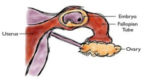
It is the process in which the fertilized egg, or a blastocyst, gets attached to the endometrial wall of the uterus to attain pregnancy. During this stage, the embryo consequently, establishes a strong connection with the maternal circulation and starts its embryonic developmental journey.
The endometrium is receptive to a fertilized egg for a shorter period in the luteal phase, which is termed as “Implantation window”. This window lasts for 4 days and extends up to 6 days after the peak of LH levels. Therefore, it is essential for the blastocyst to reach the endometrium during the implantation window in order to get a successful implantation. However, any delay in the blastocyst transport can cause loss of endometrial receptivity. However, blastocyst implantation occurs 6-8 days after fertilization.
Embryo Implantation process is mainly divided into three stages:
1. Apposition:
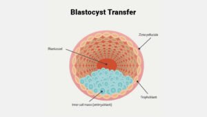
In this stage, the blastocyst sheds its zona and approaches the endometrium with a particular orientation. Meanwhile, the finger-like projections known as chorionic villi help to bring the blastocyst nearer to the uterine epithelium. Furthermore, the uterine epithelium cells have glycoprotein on their surface which acts as a barrier between blastocyst and endometrium. To overcome this barrier, the blastocyst uses its enzymes to digest the glycoprotein, ultimately resulting in the interlinking of the trophoectoderm with the luminal epithelial cells.
2. Adhesion:
Attachment occurs on the 6th or 7th day post-ovulation. At this stage, a strong association is established between the endometrium and blastocyst. Due to enzymatic breakage of glycoprotein, the adhesion molecules (includes cadherins, integrins, glycan, & lectin etc.) in blastocyst and endometrium have free access to each other.
3. Invasion:
In this stage, the embryo needs to connect with the mother for development. The blastocyst completely penetrates the endometrium by day 9. The blastocyst consists of two main components – The trophoblast & the Inner Cell Mass (ICM). Specifically, the function of trophoblast cells in the embryo is to invade the endometrium, thereby establishing a maternal connection with the embryo. Meanwhile, the inner cell mass gives rise to a fetus. As embryonic trophoblast makes contact with endometrium it further differentiates into two layers: Cytotrophoblast & Syncytiotrophoblast.
The ICM in the embryo makes two differentiated layers hypoblast & Epiblast. The cavity that arises between Epiblast & Cytotrophoblast is known as amniotic cavity. Additionally, cells originate from hypoblast which forms a thin membrane called as exocoelomic membrane. Both the hypoblast and the exocoelomic membrane contribute to the formation of the wall of the primary yolk sac. The growth of trophoblast forms Cytotrophoblast & Syncytiotrophoblast becomes quicker at this stage. The lacunar networks begin to form in the Syncytiotrophoblast & by the 12th day the lacunar networks stop growing. The uterine blood vessels in the endometrium come in contact with lacunar networks which directs the placental trophoblast to maternal blood circulation. Furthermore, the endometrial stroma becomes a dense cellular matrix called decidua, which afterwards becomes the maternal part of the placenta.
In Assisted reproduction, the key cause of implantation failure is the hatching disability of the embryo. Presently, no research is available to distinguish the embryos which can hatch and grow into a healthy embryo. No hatching is a possible reason for pregnancy failure in many elderly women. In that case, zona pellucida can be disrupted by using a laser-assisted hatching technique to improve the pregnancy rate. Reviva Fertility Clinic & IVF Center In Chandigarh offers all the latest techniques which can treat various types of infertility.
For more information about infertility please visit our website www.revivaivf.com


